Figure 4.
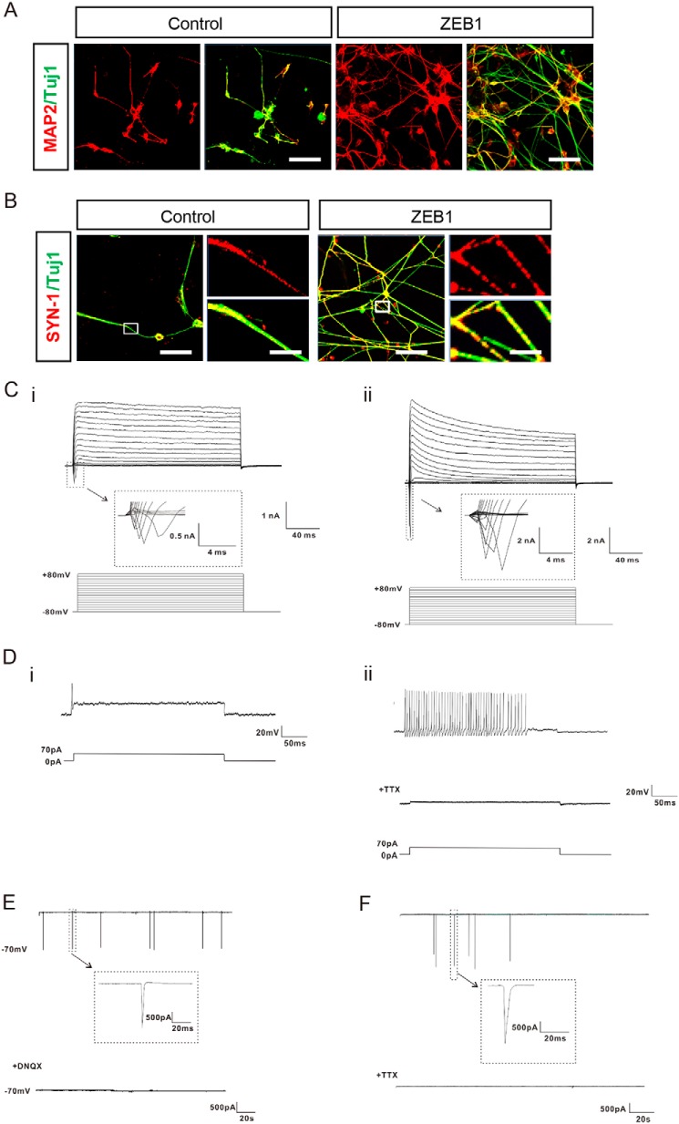
ZEB1 promoted neuronal maturation. A, immunostaining for MAP2 along with TUJ1 showed the neuronal differentiation of hESCs with or without overexpression of ZEB1 at 8 weeks of neural differentiation; n = 3. B, representative immunofluorescent images showing the localization of SYNAPSIN-1 (SYN-1) in the cells. The boxed areas at the left were enlarged as shown in the right panel, respectively; n = 3. Scale bar: 50 m (each left panel) and 5 μm (each enlarged right panel). C–E, whole cell patch recordings indicated that neurons derived from hESCs after 8 weeks of differentiation were electrophysiological active; n = 3. C, representative traces of whole cell currents were elicited by stepwise depolarizations from −80 to 80 mV from a holding potential of −70 mV in neurons of (i) control and (ii) ZEB1 group, respectively. The inset shows sodium currents. D, representative traces of action potentials (APs) from a 70 pA current injection were shown for neurons of control (i) and the ZEB1 group (ii), respectively. (i), APs in control neurons exhibited single or typically diminished amplitude in peak value. (ii), repetitive trains of APs on the similar spikes were detected in ZEB1-overexpressed neurons from neural differentiation. The fired APs could be blocked completely by TTX (1 μm). E, representative voltage-clamp recordings of spontaneous postsynaptic currents (PSCs) from neurons of the ZEB1 group after 8 weeks of differentiation. The GluR currents could be blocked with DNQX (20 μm). Cells were held at −70 mV. F, after 10 weeks of neural differentiation without forced expression of ZEB1, spontaneous PSCs held at −70 mV were detected in the neurons from the control group, and the synaptic activity could be blocked by TTX (1 μm).

