Figure 6.

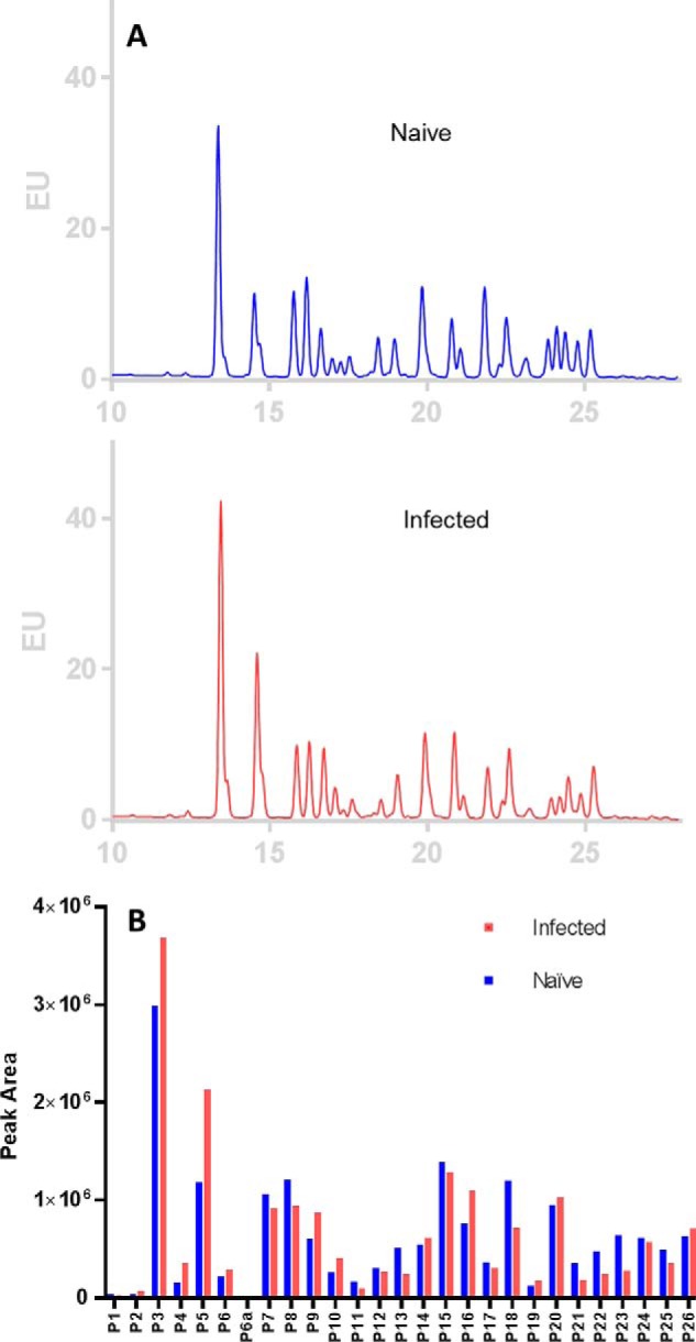
Quantification and comparison of N-glycans of IgGs from naïve to infected ferrets determined by HILIC UPLC fluorescence. A, N-glycans released from IgGs purified from pooled sera of four ferrets before (naïve) and after influenza A/H3 infection (infected). N-glycans were labeled by RapiFluor-MS and separated on HILIC UPLC monitored by fluorescence, and their chromatograms were overlaid. B, the relative abundance of corresponding peaks of N-glycans of IgGs from naïve and infected samples was compared.
