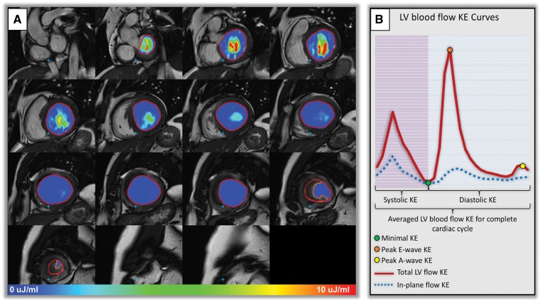
Figure 2.
(A) Left ventricular short-axis endocardial segmentation in patient with LV thrombus. Intra-cavity thrombus was manually contoured (orange contour) to avoid under-estimation of LV KE parameters. Intra-cavity KE of blood is demonstrated at peak late ventricular filling (peak A-wave). (B) Illustration of KE curve demonstrating majority of the KE parameters studied.

