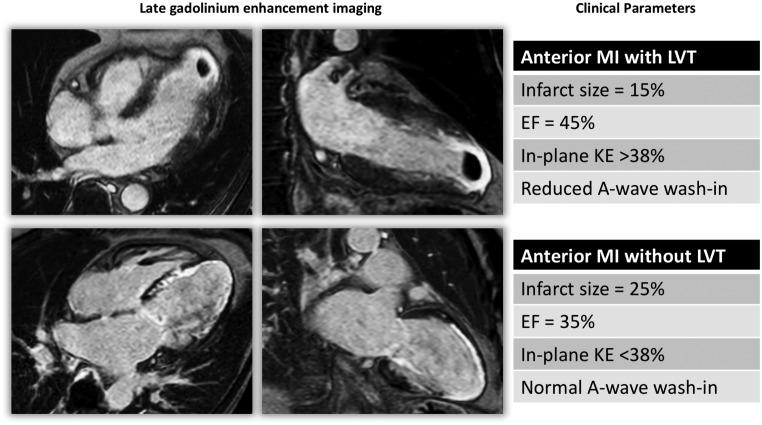
Figure 3.
Late gadolinium enhancement imaging of two case examples from the study population with anterior MI. Infarct characteristics alone did not differentiate the two cases for LVT. Left ventricular KE flow analysis demonstrated rise in in-plane, rotational KE, and reduced A-wave wash-in of the LV. EF, ejection fraction; KE, kinetic energy; LVT, left ventricular thrombus.

