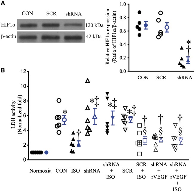
Figure 7.
Hypoxia-induced factor 1α (HIF1α) knockdown blocked isoflurane (ISO)-induced decrease in lactate dehydrogenase (LDH) activity in co-cultured endothelial cells (ECs) and cardiomyocytes (CMs) subjected to hypoxia/reoxygenation injury. (A) The expression of HIF1α protein in ECs treated with lentiviral vector containing either HIF1α shRNA (shRNA) or scrambled sequence (SCR) (left: representative western blot bands of HIF1α from the extract of cultured ECs). *P < 0.05 vs. CON (control); †P < 0.05 vs. SCR (n = 5). (B) LDH activity in media released from co-cultured ECs and CMs. All cells were subjected to 2 h of hypoxia followed by 2 h of reoxygenation (CON); whereas the cells in normoxia group underwent all culture procedures without hypoxia/reoxygenation. The values of the control groups were arbitrarily defined as one in each batch of experiments. Data are presented as mean ± SEM. Kruskal–Wallis test followed by Dunn’s test was used to analyse multiple group comparisons. *P < 0.05 vs. normoxia; †P < 0.05 vs. CON; ‡P < 0.05 vs. ISO; §P < 0.05 vs. shRNA (n = 6). rVEGF, recombinant vascular growth factor.

