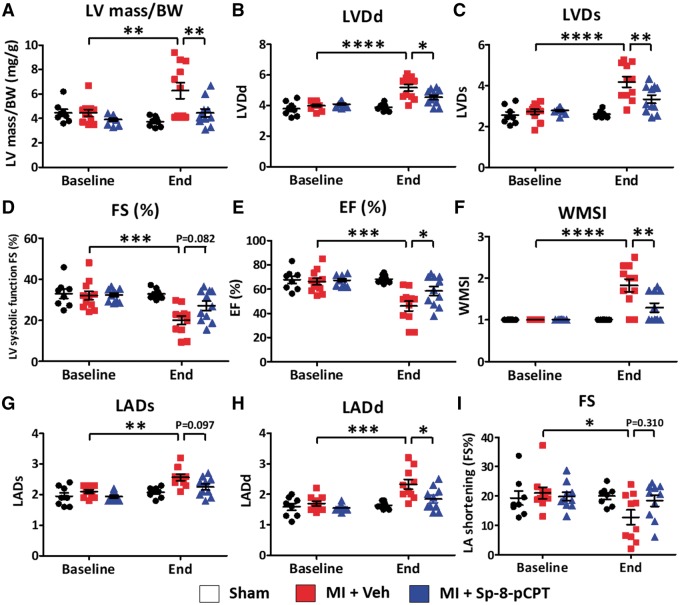
Figure 7.
Effect of Sp-8-pCPT on cardiac function. Echocardiography was performed at baseline and 2 weeks post-myocardial infarction (MI). (A) Ratio of left ventricular mass to body weight (BW) was increased 2 weeks after MI; this increase was prevented by Sp-8-pCPT. (B–H) Sp-8-pCPT prevented MI-induced impairment in LV systolic function, LV dilation, left-atrial dilation, and wall-motion abnormalities. (I) Sp-8-pCPT did not change LA shortening. (*P < 0.05, **P < 0.01, ***P < 0.001, ****P < 0.0001, two-way ANOVA followed by Tukey’s test, n = 7–11 mice/group.) Each dot represents an individual animal. LVDd, left ventricular dimension at end-diastole; LVDs, left ventricular dimension at end-systole; FS, fractional shortening; EF, ejection fraction; WMSI, wall motion score index; LADs, left atrial dimension at the end of ventricular systole; LADd left atrium dimension at the end of ventricular diastole.

