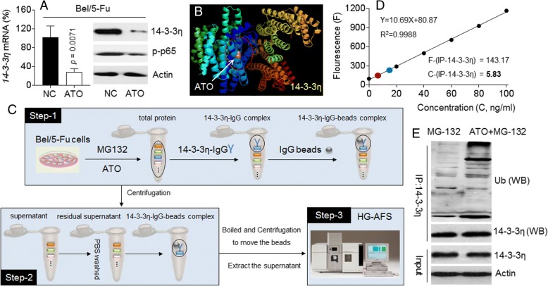
Fig. 6.
Relationships between ATO and 14–3-3η: a Bel/5-Fu cells were treated by 10 μM of ATO for 24 h. qRT-PCT analyses in triplicate (left) and Western blot (right) analyses of the expressions of 14–3-3η and/or p-p65. b PyMol software analyses of the binding of ATO to 14–3-3η. cThe procedure of IP-AFS. d and e After Bel/5-Fu cells were pre-treated by 20 μM of MG-132 for 2 h, they were exposed to 10 μM of ATO for 6 h. The 14–3-3η was immunoprecipitated with the specific antibody. d AFS analyses of the concentration of ATO in 14–3-3η-immunoprecipitation complex. e Western blot analyses of the expression of ubiquitin in 14–3-3η-immunoprecipitation complex

