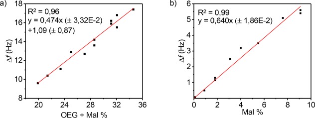
Figure 3.
(a) QCM-D frequency shift (fifth overtone) of the PLL-OEG-Mal deposition step versus the total degree of functionalization of the modified PLL, quantified by 1H NMR. (b) QCM-D frequency shift of the cDNA binding step versus the fraction of Mal grafted to the PLL polymer, quantified by 1H NMR. All experiments were performed using 0.3 mg/mL of modified PLL, 1 μM PNA thiol solution (activated by TCEP), and 1 μM cDNA solution in PBS at pH 7.2. PLL-OEG-Mal polymers with different degrees of functionalization were used (Mal = 0.0–9.1%, OEG 15.9–29.1%). Data points represent individual measurements.

