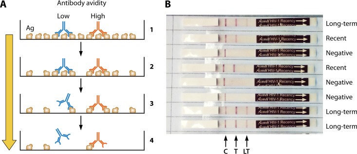
FIG 5.
Schematic representations of the assay principle for a limiting-antigen (LAg) assay to differentiate low- and high-avidity antibodies (A) and a rapid incidence-prevalence assay (B). (A) At high antigen concentrations, bivalent antibodies of both low avidity (blue) and high avidity (red) bind to microwells coated with antigen, whereas at limiting antigen concentrations, only the high-avidity antibodies bind due to monovalent binding and are retained after washing. (B) Two empirically optimized antigen concentrations are incorporated onto the nitrocellulose strip for diagnosis of HIV infection and detection of recent infection. C, control line; T, test line at a high antigen concentration; LT, low antigen concentration to differentiate recent from long-term infections. The binding of antibody is determined with lateral flow technology. The control band indicated by the arrow is an internal IgG procedural control. Seronegative samples are reactive only for this control band. Persons with recent infections will show C and T lines but not the LT line, and those with long-term infections (>1 year) are reactive to all three lines, including the LT line.

