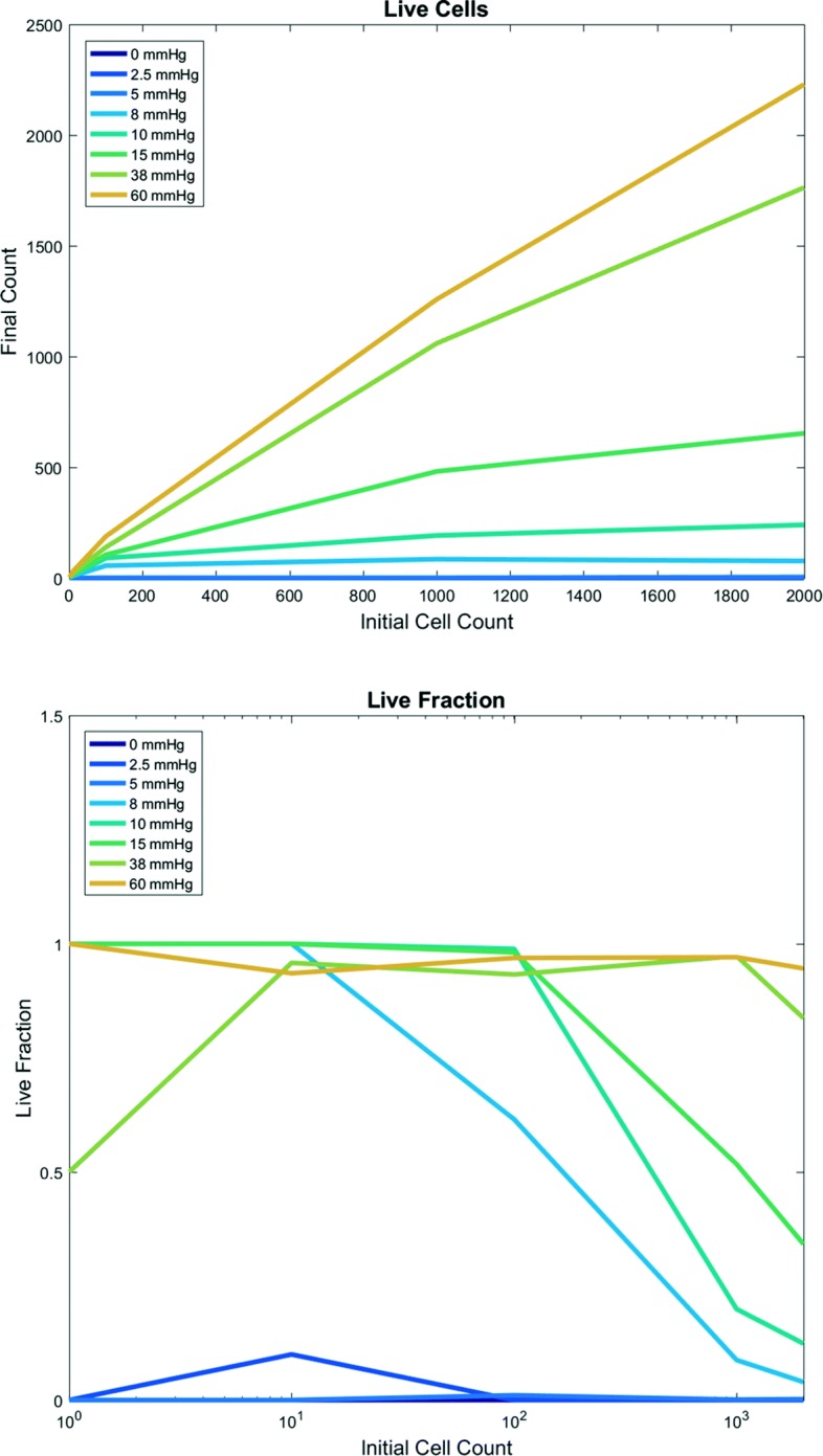
Fig. 4.

First PhysiCell-EMEWS test on cancer hypoxia: analytics. Live tumor cell count (top) and live cell fraction (bottom) after 48 h, as a function of oxygenation conditions (each curve is a different condition) and initial cell count (horizontal axis). For intermediate oxygenation conditions, increasing the initial cell count increases the final live cell count (top) but decreases the live cell fraction (bottom). Once oxygenation is high enough, any initial cell count yields nearly 100% live fraction at 48 h
