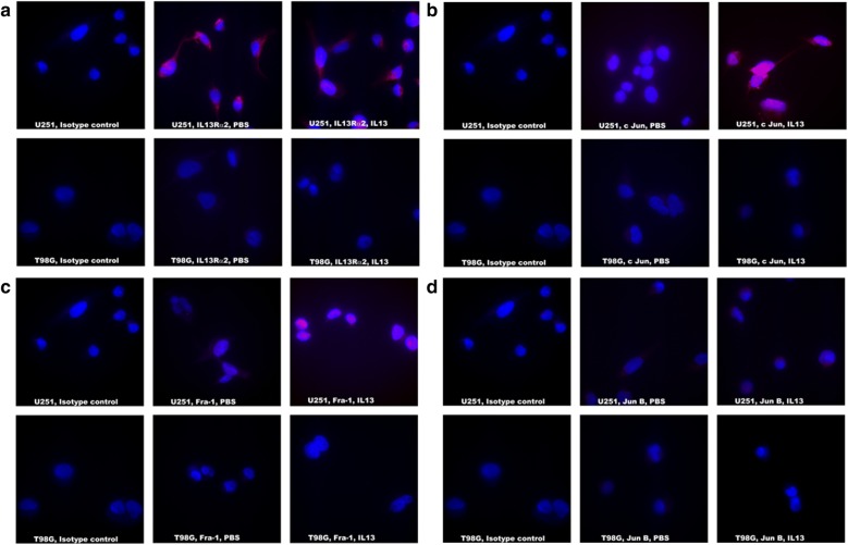
Fig. 1.
Evaluation of AP-1 transcription factors in IL-13 treated U251 and T98G cell lines. a IL13Rα2 staining with Streptavidin 594 (red) and DAPI used for nuclear staining (blue). b c Jun staining with Streptavidin 594 (red) and DAPI used for nuclear staining (blue). c Fra-1 staining with Streptavidin 594 (red) and DAPI used for nuclear staining (blue). d Jun B staining with Streptavidin 594 (red) and DAPI used for nuclear staining (blue). All images were captured at ×1000 magnification

