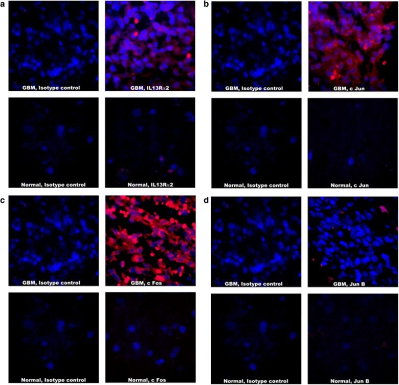
Fig. 4.
Analysis of AP-1 transcription factors in Human GBM samples and normal human brain sample. a IL13Rα2 staining with Streptavidin 594 (red) and DAPI used for nuclear staining (blue). b c Jun staining with Streptavidin 594 (red) and DAPI used for nuclear staining (blue). c c Fos staining with Streptavidin 594 (red) and DAPI used for nuclear staining (blue). d Jun B staining with Streptavidin 594 (red) and DAPI used for nuclear staining (blue). Immunostained sections were photographed at ×1000 magnification. Extent of immunostaining and percent positive field were counted and analyzed

