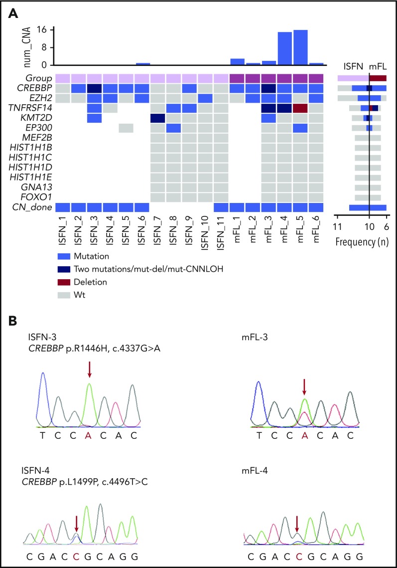
Figure 1.
Mutation overview in mFL and ISFN cases. (A) The heat map shows the case-specific pattern of mutations found by Sanger sequencing and NGS. Each column represents a case sorted by ISFN and mFL. Each row represents a gene. (Right) Bar graph illustrates the mutation frequency of each gene in a specific entity. (Top) Bar graph indicates the number of copy number alterations detected by array-comparative genomic hybridization in a previous study.13 Loss of heterozygosity determination based on polymorphic single-nucleotide polymorphism (data not shown) indicated TNFRSF14 CNN-LOH alteration in mFL-3. mFL cases 1 and 2 were analyzed for mutations in CREBBP and EZH2 with Sanger sequencing only. (B) Examples of CREBBP mutations detected in 2 paired ISFN/mFL samples by Sanger sequencing. The peak of the ancestral base is higher than in the mFL case in the ISFN. The analysis was performed in microdissected ISFN. CNN-LOH, copy number neutral loss of heterozygosity; wt, wild-type.

