Fig. 6.
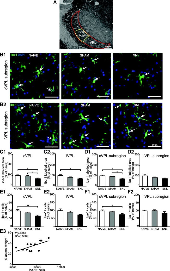
Decreased iba-1 immunostaining and decreased number of iba-1-positive cells in the contralateral VPL 14 days after spinal nerve ligation. Panel A illustrates the delineation of the ventral posterolateral thalamic nucleus (VPL; outlined in red) with its subregion that receives information from the hind limb (VPL subregion; outlined in yellow) on a rat brain section treated for visualizing acetylcholinesterase activity. The outlines were then applied on an adjacent section treated for the immunohistofluorescent detection of the iba1. Microphotographs in B1 and B2 are examples of iba-1 immunostaining (green) and DAPI staining (deep blue) in the contralateral (cVPL, B1) and ipsilateral (iVPL, B2) subregion of the VPL in naïve, sham, and SNL animals 14 days after the beginning of the experiments. White arrows point at iba-1/DAPI-positive cells (part of the cell body appears in turquoise, that is to say mix of green and deep blue). Note that there is less iba-1 immunostaining and less iba-1/DAPI-positive cells in the cVPL subregion of SNL rats. Morphometric analysis conducted on the VPL (C1 and C2) and on the VPL subregion (D1 and D2) reveals that iba-1 immunofluorescent surfaces (expressed as percentage of naïve values) are significantly decreased in the cVPL (C1: one-way ANOVA F(2, 13) = 5.151, p = 0.0225) as well as in the cVPL sub-region (D1: one-way ANOVA F(2, 13) = 4.716, p = 0.0288) in the SNL rats (n = 6, 3 slices per animal) compared to naïve (n = 4, 3 slices per animal) and sham (n = 6, 3 slices per animal) rats. This decrease is also found in on the ipsilateral side (C2 and D2) but only reaches statistical significance in the iVPL of SNL rats compared to naïve rats (C2: one-way ANOVA F(2, 13) = 4.183, p = 0.0396). Quantification of iba-1/DAPI-positive cells in the VPL (E1 and E2; cell number per volume unit expressed as percentage of naïve values) by using an optical disector method of stereological counting and in the VPL subregion by using a conventional method (F1 and F2; cell number per surface area unit expressed as percentage of naïve values) reveals that the number of iba-1/DAPI-positive cells is significantly reduced in the cVPL (E1: one-way ANOVA F(2, 13) = 6.669, p = 0.0102) as well as in the cVPL subregion (F1: one-way ANOVA F(2, 13) = 4.679, p = 0.0295) of SNL rats compared to the one of naïve rats. No significant difference is found in the iVPL (E2: one-way ANOVA F(2, 13) = 3.784, p = 0.0507) and the iVPL subregion (F2: one-way ANOVA F(2, 13) = 2.878, p = 0.0923). The number of iba-1/DAPI-positive cells in the cVPL is correlated to the ambulatory pain. The less weight the animal bears on its ipsilateral hind paw, the lower the number of iba-1/DAPI positive cells is (E3). Post hoc test (Tukey’s multiple comparison test): *p < 0.05; **p < 0.01

