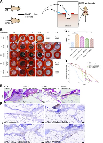
Figure 6.
siKeap1-transfected dBMSCs reconstitute role in repair. A: BMSCs from WT or db/db mice were cultured and prepared 24 h prior, with or without transfection with siKeap1. Twenty-four hours postexcision, 5,000,000 cells in 300 μL were injected into the cutaneous wound bed and immediate periphery. The wound bed was seeded with 100 μL, and the remaining 200 μL was seeded in 50-μL aliquots in the immediate wound periphery. B: Photographs of excisional wounds inoculated with BMSCs as indicated. C: Mean time to closure of cutaneous wounds. D: Photometric quantification of wound area. E: Hematoxylin-eosin stains of tissue sections of 10-day wounds. Wound is to the left in the image. Dashed black line, wound edge epidermis. Yellow lines delineate granulation tissue area. F: CD31 immunohistochemistry on 10-day wound tissue. Scale bars, 100 μm. Data represented as mean ± SD; n = 4. **P < 0.01. See also Supplementary Fig. 5.

