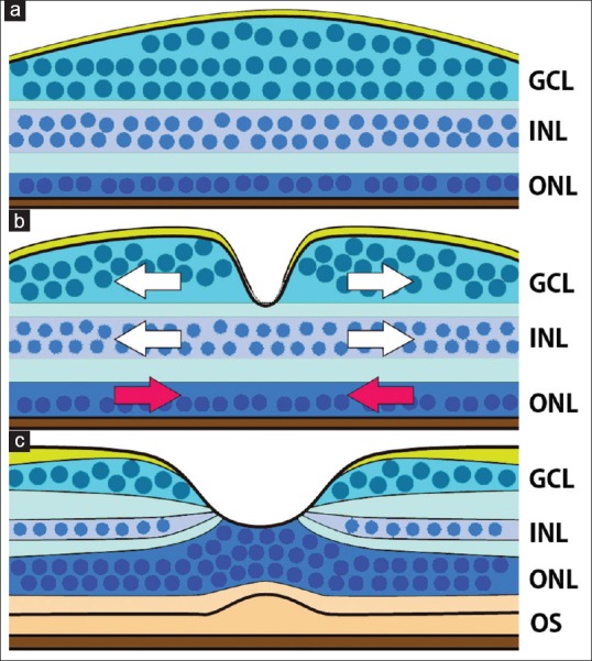
Figure 1.

Schematic diagram showing the development of the foveal structure. (a) At the end of the second trimester, a thickened ganglion cell layer is found, however, a foveal pit is not present. The outer nuclear layer is a single-cell layer. (b) A foveal pit forms in the ganglion cell layer and inner nuclear layer. The cells in the ganglion cell layer and inner nuclear layer are displaced centrifugally to form the foveal pit (white arrows), and cone cells in the outer nuclear layer are displace centripetally (red arrows). (c) In the adult retina, the cone cells form a multicellular layer, and the inner and outer segments become prominent. The schema is based on references Springer and Hendrickson [23] and Hendrickson[29]
