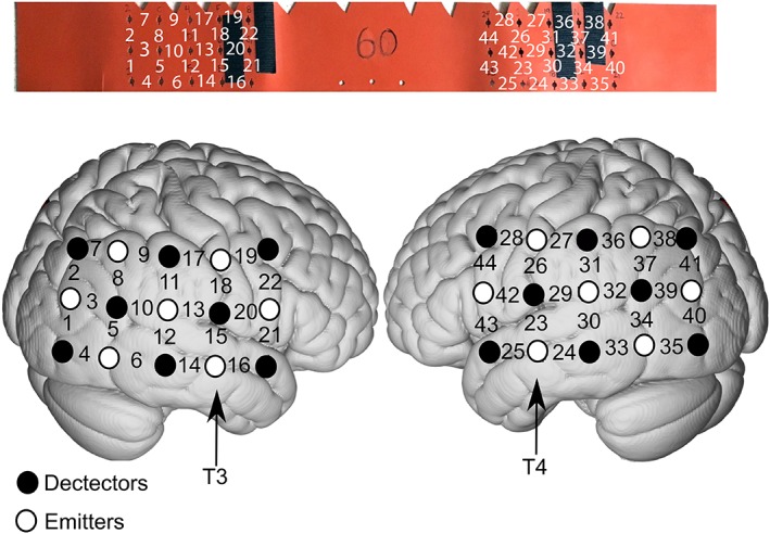
Figure 2.

Example of cap configuration for auditory research based on the 10‐20 EEG system. T3 and T4 are used as anatomic references when placing the cap. For this design, channels 13 and 15 and channels 23 and 29 record from primary auditory cortex and surrounding belt regions of the right and left hemisphere, respectively. The top panel shows the cap/band used when a subject's head circumference measures 60 centimeters. The bottom panel is a schematic of the cortical regions associated with each optode and associated brain region.
