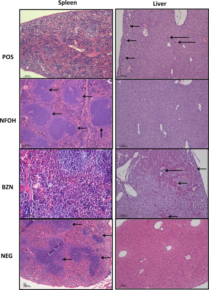
Figure 8.

Photograph of the spleen and liver tissues (H&E, 10×). Spleen: POS and BZN lymphoid follicles unorganized, NFOH and NEG lymphoid follicles organized (small arrow); liver: POS and BZN presence of inflammatory infiltrates (larger arrow) and necrosis areas (small arrow), NFOH and NEG healthy hepatocytes [Colour figure can be viewed at wileyonlinelibrary.com]
