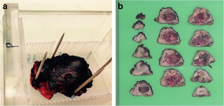
Fig. 1.
(a) Specimen I containing a 20-mm large oncocytoma, fixated on a block of paraffin inside a customised Perspex holder with a Perspex row of pins on both sides 3 mm apart, used for pathology slicing after fixation in formalin. The holder also contains seven (two posterior left) water-filled tubes (blue pins) to facilitate matching between MR imaging and histopathology slides. The total setup is positioned in a glass container. (b) After MR examination the oil in the glass container was disposed of and the specimen was fixated at least 24 h in formalin. Subsequently, the specimen was cut in 3- to 5-mm thick whole mount sections from anterior to posterior using the pins in the holder and totally included for pathology work-up

