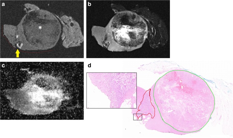
Fig. 2.
(a) Specimen X containing a 24-mm large clear cell tumour and a 10-mm large incidentally detected papillary tumour. The T1-weighted images show the clear cell (*) and papillary tumour (#), the surgical margin (red line), and suspected positive surgical margin (yellow arrow.) Image quality was scored as ‘3 – acceptable’. (b) According to T2-weighted images. Image quality was scored as ‘1 – excellent’. (c) According to calculated ADC map. Image quality was scored as ‘4 – poor’. (d) Histopathological slide with enlargement shows demarcation of the clear cell (green) and papillary (red) tumour. The enlargement does not confirm tumour cells in the resection border

