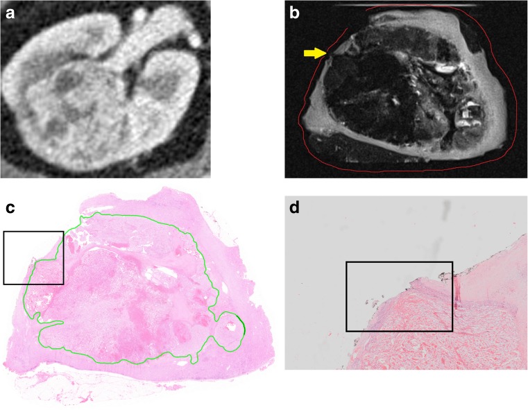
Fig. 3.
(a) Preoperative CT scan of specimen III showing a 50-mm large clear cell RCC in the right kidney. (b) The T2-weighted scan of the specimen after resection showed suspicion for a positive (yellow arrow) surgical margin (red line). (c) Histopathological slide confirmed the 1.4-mm large positive surgical margin. Black box is enlarged in Fig. 3d. (d) The 1.4-mm large positive surgical margin in detail

