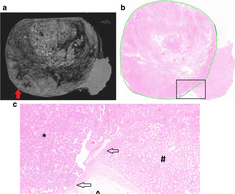
Fig. 4.
(a) Specimen VIII containing a 70-mm papillary tumour showing a false-positive result for presence of a pseudocapsule (red arrow) on the T1-weighted scan; the specimen is slightly compressed at the top to fit in the setup. (b) The annotated histopathological slide shows the tumour borders (green line). The black square is enlarged in c. (c) The structure marked as a pseudocapsule on MRI was found to be a thin epithelial layer surrounding the tumour mimicking a pseudocapsule. Markers are as follows: * tumour tissue; # renal parenchyma, ^ adipose tissue; arrows mark the epithelial layer

