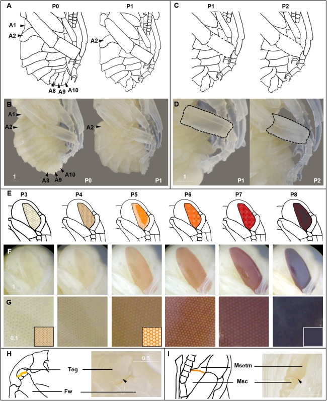
Figure 3. Morphological criteria marking transitions to P0–P8.
(A & B) Illustration and photo for transition from P0 to P1. Note the retraction of A1 and A8–A10. (C & D) Illustration and photo for changes in shapes of basitarsi of the hind leg (broken lines) of B. impatiens workers in the transition from P1 to P2. (E–G) Illustration (E) and photo (F) for color shift of the CE from P3 to P8. (G) shows zoomed view of the CE revealing appearance of ommatidial units at different stages, including dotted (P3 and P4), then hexagonal (P5–P7) and finally, a filled pattern (P8-eclosion). (H) and (I) Black triangles indicate a slightly tanned suture at forewing-tegula junction (H) at P3, and between the mesepisternum and mesocoxa (I) at P7. Teg, tegula; Fw, forewing; Msetm, mesepisternum; Msc, meso-coxa. Scale bar unit: mm.

