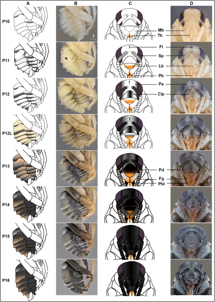
Figure 5. Morphological criteria marking the transition to P10–P16.
(A & B) Photo and schematic illustration of melanization patterns of abdominal cuticle. Melanization first appears as peppered stripes on the apical margin of each metasomal tergite/sternite disc (P10–P12L), then expands anteriorly to cover the whole disc (P13–P16). The orange tinge at P12L/P13 demonstrates the beginning of tanning of the cuticle. Orange at P14–P16 indicates partial pigmentation of black setae, while the white in T2 (P14–P16) indicates the white color of the future yellow hairs, as the underlying cuticle at these stages is mostly or all black. (C & D) Photo and schematic illustration of pigmentation on the head region including the face, mouthparts and antennae. Mb, mandible; Th, teeth; Fl, frontal line; Sp, Scape; Lb, labrum; Pb, proboscis; Pa, paraocular area; Clp, clypeus; Pd, pedicle; Fg, flagellomeres; Pbl, lateral-basal proboscis. Black triangle: broken melanization stripes at stage P11. Scale bar unit: mm.

