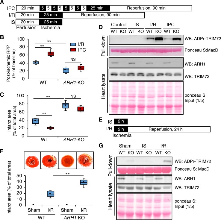
Figure 3. Ischemia/reperfusion injury enhanced ADP-ribosylation of TRIM72 in ARH1-KO mouse heart in vivo and in perfused heart model.
(A) Langendorff isolated perfused heart model protocol. IPC, ischemic preconditioning; I/R, ischemia/reperfusion; IS, ischemia. (B and C) Recovery of postischemic left ventricular (postischemic rate pressure product [RPP]) (B) and myocardial infarct size (C) after I/R or IPC in ARH1-KO mice (male, n = 6, 33.9 ± 0.8 weeks, n = 5, 34.5 ± 1.3 weeks; female, n = 6, 34.8 ± 0.4 weeks, n = 5, 33.9 ± 1.9, respectively) and in WT mice (male, n = 4, 33.6 ± 1.3 weeks, n = 5, 37.9 ± 1.8; female, n = 4, 34.4 ± 0.6 weeks, n = 5, 38.5 ± 2.9, respectively). (D) ADP-ribosylated TRIM72 collected by Af1521 macrodomain–GST pull-down assay (n = 3). (E) Myocardial ischemia/reperfusion protocol in vivo. (F) Images of TTC stain and measurement of infarct size after sham operation and in vivo I/R in ARH1-KO (n = 6, n = 7, respectively) and WT (n = 7, n = 7, respectively) mice. (G) Endogenous ADP-ribosylated TRIM72 increased after I/R in ARH1-KO but not WT mice. ADP-ribosylated TRIM72 was bound to immobilized Af1521 macrodomain–GST (Af1521). ADPr-TRIM72 coimmunoprecipitated with Af1521 macrodomain–GST was detected by Western blotting (WB) with anti-TRIM72 antibody. Box-and-whisker plots show median, first and third quartiles (boxes), and minimum and maximum values (whiskers). **P < 0.01, by 1-way ANOVA, post hoc Student-Newman-Keuls test. Immunoblot data are representative of 3 experiments.

