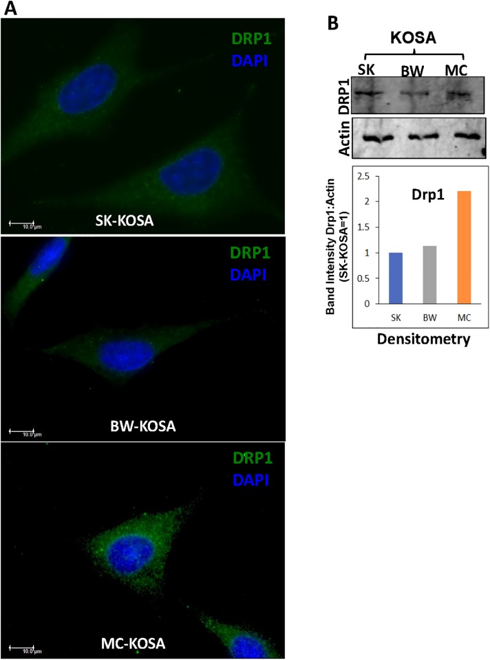
Fig 3. Altered expression of mitochondrial fission marker in OSA cell lines.
(A) Immunocytochemistry of OSA cell lines stained for mitochondrial fission marker protein Drp1 (green) and nuclei (DAPI, blue) viewed under a Leica widefield microscope. Scale bar: 10μm, magnification 100x. (B) Top panel: Western Immunoblot showing high DRP1 protein levels in MC-KOSA relative to SK-KOSA and BW-KOSA. Bottom Panel: Quantitation (densitometry) of the protein levels from the western immunoblot.

