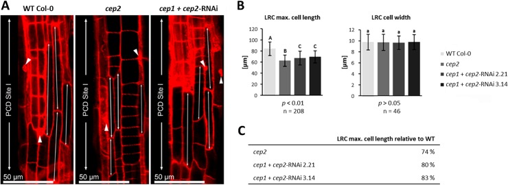
Fig 4. CEP2 loss of function results in shortening the longest cells of the lateral root cap (LRC) at the level of the PCD site I.
The residual CEP2 activity in cep1 cep2 double ko/kd mutants (cep1+cep2-RNAi) results in prolonged LRC cells as compared to cep2 ko mutants, but shorter as compared to LRC cells in WT plants. (A) 7 days old seedlings of WT, cep2 ko and cep1 cep2 double ko/kd mutants (cep1+cep2-RNAi) were stained with propidium iodide and analyzed by CLSM (single pictures, 400-fold magnification). The 3–5 longer most cells of the LRC at the level of the PCD site I (double arrow) are compared. Nuclei stained with propidium iodide are marked (arrow heads) indicating cells approaching PCD since their cell membranes become permeable resulting in propidium iodide stained nuclei in addition to the stained cell wall. (B) Comparison of the length (left) and the width (right) of the 3–5 longer most LRC cells in 7 days old seedlings of WT plants with cep2 ko and cep1 cep2 double ko/kd mutants (cep1+cep2-RNAi; lines 2.21 and 3.14) mutants. Columns are marked with different letters indicating statistically different groups for LRC cell length (p<0.001) or with similar letters indicating groups not statistically different for LRC cell width (p>0.05) according to the ANOVA-and Duncan test. Data represent the respective means of 208 plants in three independent experiments (biological replica) each comprising 15 seedlings per line (experiment 1 and 2, each) or 16 plants (experiment 3) per genotype resulting in 208 longer most LRC cells (3–5 cells per LRC) evaluated for the cell length and 46 LRC cells evaluated for the cell width. (C) LRC cell length expressed as percentage of WT plants.

