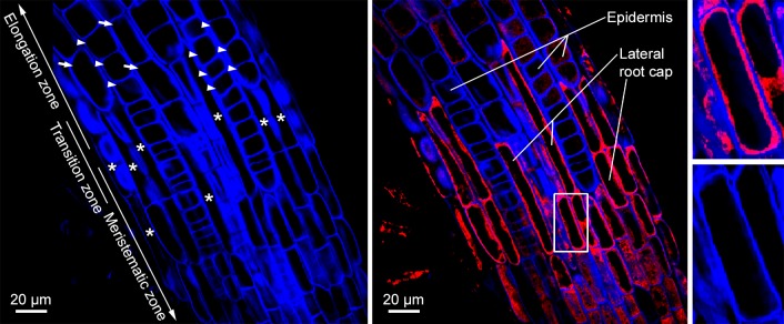
Fig 8. Pro-CEP2 accumulates within the protoplasm of the longer most cells from the upper end of the LR cap (PCD site I) at the level of the transition zone and is still present in root cap corpses.
Roots of 7 days old WT seedlings expressing the pro-mCherry-CEP2 (PCEP2::pre-pro-3xHA-mCherry-AtCEP2-KDEL) are stained with calcofluor-white in order to visualize the cell wall and analyzed by CLSM (stack of 4 optical sections, Olympus Fluoview FV 1000). Left: Visualization of cell walls (blue): isodiametric, not elongated epidermis cells (arrow heads) from the meristematic and transition zone as well as already elongating epidermis cells (arrows) from the rapid elongation zone in addition to small, elongated and already collapsing cells at the upper end of the LRC (asterisk) are found. Middle: Merge of calcofluor (blue) and pro-mCherry-CEP2 (red): pro-CEP2 is found in the protoplasm of LR cap cells and is still present in root cap corpses sticking to epidermis cells of the rapid elongation zone. Pro-CEP is localized within the cell in the protoplasm surrounding the large vacuole and not in the cell wall. Right: magnification of the white boxed area presenting a LR cap cell with the blue coloured cell wall, the large black looking vacuole and in between the protoplasm with the red coloured pro-CEP2.

