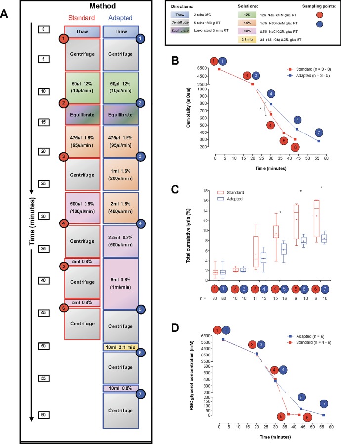
Fig 2. Comparison of the standard and our adapted deglycerolization procedure.
(A) Diagram of the standard deglycerolization protocol and our adapted method, designed to reduce RBC lysis. Samples (40% v/v glycerol final) were thawed (2min, 37°C), then centrifuged (1,500g, 5min, 4°C). For equilibration, samples were left undisturbed for 3min at RT. Circled numbers identify sampling points for the graphs presented in B-D. (B) Sample osmolality was measured at various stages of the standard and adapted deglycerolization methods. The adapted method slowed osmolality reduction during de-gycerolization, thereby protecting cells from lysis (n = 3–8 samples from 3 individual blood donors). (C) Total cumulative RBC lysis measured at different stages of the standard and adapted deglycerolization methods. RBC lysis was significantly reduced using the adapted deglycerolization method (n = samples, shown on graph. Minimum number of individual blood donors = 4). (D) Intracellular RBC glycerol measured at different stages of the standard and adapted deglycerolization methods. Both methods result in total glycerol removal (> 99.9%) (n = 4–6 samples from 3 individual blood donors).

