Fig. 3.
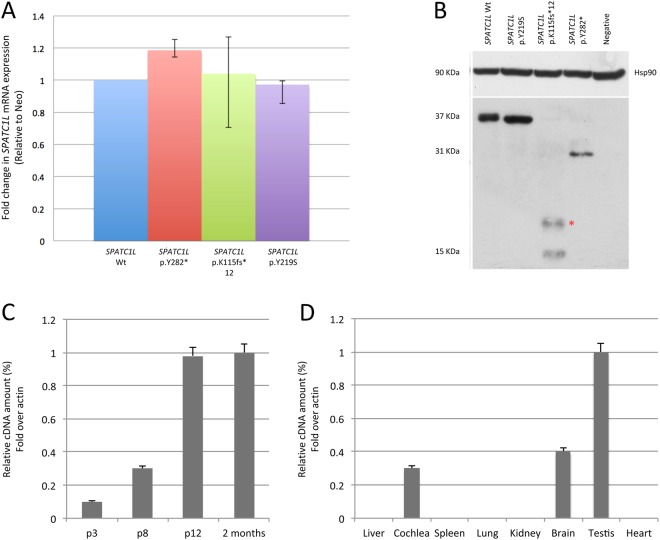
SPATC1L mRNA and protein levels in Hek293 transfected cells and Spatc1l expression in mouse whole cochlea and other tissues at different time points. a qRT-PCR analysis of relative mRNA expression of SPATC1L wild type and mutants after 48 h of transfection in Hek293 cells. Results are expressed as a fold-change of expression levels, and are normalized to the relative amount of the internal standard Neo. Error bars indicate 95% confidence intervals. b Western blot analysis of SPATC1L wild type and mutant proteins. Hsp90 was applied to determine equal loading. c The graph shows expression of Spatc1l in mouse whole cochlea at P3, P8, P12 and 2 months. Results are reported as fold change in gene expression over β actin, used as an internal control. The gene shows an age-related expression. d The graph shows Spatc1l expression at 2 months of age in different mouse tissues, including liver, cochlea, spleen, lung, kidney, brain, testis and heart. Results are reported as fold change in gene expression over β actin, used as an internal control

