Figure 7.
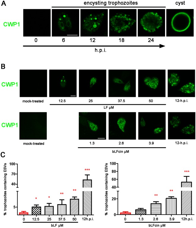
Addition of bLF and bLFcin to growing wild-type trophozoites induces CWP1 expression and ESVs formation. (A) The encystation process: Immunofluorescence assays and confocal microscopy show that CWP1 (in green) is not expressed on non-encysting trophozoites but appeared in de novo formed ESVs after 6 hours post-induction (h.p.i.) of encystation. Finally, CWP1 is released to constitute the cyst wall (cyst). Bar, 5 μm. (B) After treatment, CWP1 is observed in trophozoites exposed to different concentrations bLF or bLFcin in Labeling buffer lacking serum for 30 min. No CWP1 signal is detected in mock-treated growing trophozoites. Trophozoites encysting for 12 h (12 h.p.i) are shown as control of the IFA. Bar, 5 μm. (C) The graphics show the percentage of trophozoites with formed ESVs which increases as we raise the dose of bLF or bLFcin. Data shown are representative of three independent experiments and are expressed as means of triplicates ***p-value < 0.001, **p-value < 0.01, and *p-value < 0.05.

