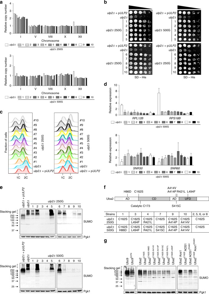
Fig. 5.
UBA2 and AOS1 genes are mutated in high-passage ulp2Δ cells. a–e qPCR ploidy assays (a), growth assays (b), flow cytometry analysis (c), qRT-PCR analysis of ribosomal protein genes RPL12B and RPS18B and snoRNA genes SNR30 or SNR60 (d), and immunoblot analysis of SUMO-conjugated proteins (e) were performed for ten independently evolved ulp2Δ lines at 250 or 500 generations. Gene expression was measured relative to WT cells (MHY1379) and data were normalized to SPT15 expression in d. The error bars indicate the SD from two experiments. f Schematic of Uba2 protein and list of mutations in ulp2Δ 250G or 500G strains. AD, CD, and UFD indicate adenylation, Cys and ubiquitin-like fold domains, respectively. Positions of the catalytic Cys residue and mutations identified in Table 2 are noted. g ubc9Δ, uba2Δ, or uba2Δ aos1Δ strains carrying a YCplac33 plasmid with UBC9, UBA2, or UBA2 AOS1 were transformed with a pRS315 plasmid expressing WT or the indicated mutant proteins. After shuffling out the URA3-marked plasmids, anti-SUMO immunoblot analysis of cell extracts was performed

