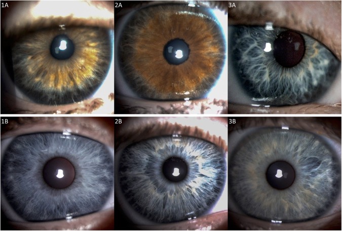
Figure 2.
Peripherally thinned irides in children with Down syndrome (Row A) versus controls (Row B). (Row A) Extensive peripheral iris thinning and absence of contraction furrows in light brown, hazel, and blue irides in children with Down syndrome. Brushfield spots were located between normal and thinned iris. (Row B) Less extensive iris thinning in control children with blue irides, which may not be present in those with darker irides (not shown). (1B,3B) Contraction furrows are present in two children, as are Wölfflin nodules, found at the demarcation between normal and thinned iris.

