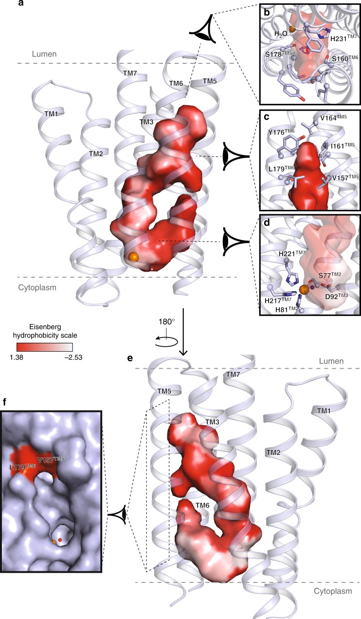
Fig. 2.
ACER3 intramembrane domain. a View of the large hook-shaped internal cavity shown as surface (cavity mode 1) within the 7TM helix bundle (shown in light blue cartoon). The TM4 has been removed for clarity. The cavity is colored according to the Eisenberg hydrophobicity classification from red (high hydrophobicity) to white (low hydrophobicity). b, c Close-up views of the pocket on the top highlighting the observed density (blue mesh, 2Fo–Fc map contoured at 1σ) in which a water molecule was modeled (red sphere) b and on the side c with residues lining the pocket shown as sticks. d Close-up view of the zinc binding site highlighting the residues forming the first coordination sphere of the Zn2+ shown as sticks. The modeled water molecule is shown as a blue sphere. e 180° rotation of the view described in (a). f Side view of the pocket shown as surface (cavity mode 0) revealing the pocket accessibility at the level of the Zn2+ site and right above it

