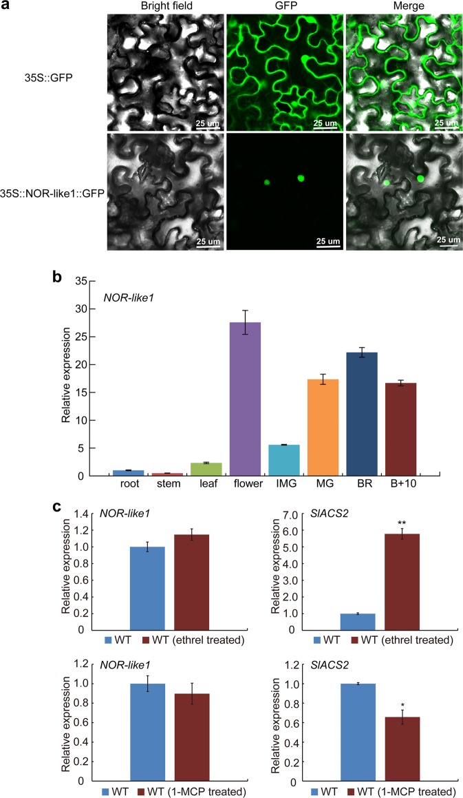
Fig. 2. Subcellular localization of NOR-like1 in nuclei, gene expression pattern of NOR-like1 and the expression of NOR-like1 in WT fruit after treatment with ethrel and 1-MCP.
a Subcellular localization of NOR-like1 in nuclei. Tobacco leaves were used for subcellular localization. Green fluorescence images were taken in a dark field, while the outline of the cell was photographed in a bright field. 35S:NOR-like1:GFP represents NOR-like1 and GFP fusion protein. 35S:GFP represents the control. Bars = 25 μm. b qRT-PCR analyses of NOR-like1 in different tomato organs and fruit ripening stages. IMG, immature green. Actin was used as the internal control. Bars represent ± SD of three independent replicates. c Expression of NOR-like1 in WT fruit after treated with ethrel and 1-MCP. Actin was used as the internal control. SlACS2 was detected as the positive control. Error bars indicate ± SD of three biological replicates. Asterisks indicate significant differences determined by Student’s t-test (**p < 0.01, *p < 0.05)

