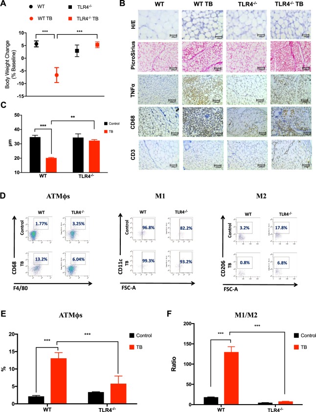
Figure 1.
TLR4 deletion attenuates scAT remodeling during cancer cachexia syndrome. (A) Wild-type C57BL/6 and TLR4−/− mice (8-week old male) were inoculated with LLC tumor cells. Body weight change (excluding tumor weight) was evaluated after 28 days before inoculated tumor cells. N = 10 per group. (B) Histologic sections of scAT in different experimental groups. Histological staining for H/E and picrosirius red were performed, and also, immunohistochemistry for inflammatory profile (TNFα) and immune cell markers (CD68 and CD3). N = 5 per group. (C) The size of adipocytes (cell diameter) from WT and TLR4−/− mice was quantitatively analyzed (500 adipocytes were measured for each group) after the experimental protocol. N = 5 per group. (D) Stromal vascular fractions (SVF) were isolated from scAT by collagenase digestion for each different group. Flow cytometric analysis of SVF was conducted using fluorescent-conjugated antibodies against CD68, F4/80, CD11c, CD206. Adipose tissue macrophages (ATMϕs) were defined as CD68+F4/80+ subpopulations and displayed the values as percentage of your respective groups. M1 and M2 ATMϕs were defined as CD68+F4/80+CD11c+CD206− and CD68+F4/80+CD11c−CD206+, respectively. Representative flow cytometric dot plots showing the percentage of ATMϕs. N = 4 per group. (E) Quantification of double-positive cells for CD68+F4/80+ related to dot plots showed in (D). (F) M1/M2 ATMϕs ratio in the scAT. Scale bars, 100 μm and 200 μm. Graphs show the mean ± SEM. Statistical significance was determined by two-way ANOVA. **P < 0.01; ***P < 0.001.

