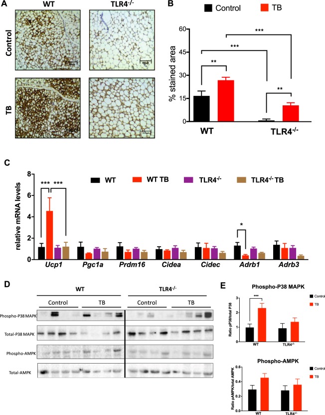
Figure 3.
TLR4 deletion reduces browning effect in TB mice throughout p38MAPK pathway. (A) Representative images of UCP1 staining of scAT from the different experimental groups. N = 5 per group. (B) Total quantification of UCP1 staining. (C) qRT-PCR was performed to quantitate Ucp1, Pgc1a, Prdm16, Cidea, Cidec, Adrb1 and Adrb3 mRNA levels in scAT from the different groups. N = 5 per group. (D) Depicted are representative immunoblots to detect phospho-p38MAPK, p38MAPK, phospho-AMPK and AMPK levels in scAT from the different experimental groups. N = 4 per group. (E) Densitometric evaluation of protein levels (phospho/total). Scale bars, 200 μm. Graphs show the mean ± SEM. Statistical significance was determined by two-way ANOVA. *P < 0.05; **P < 0.01; ***P < 0.001.

