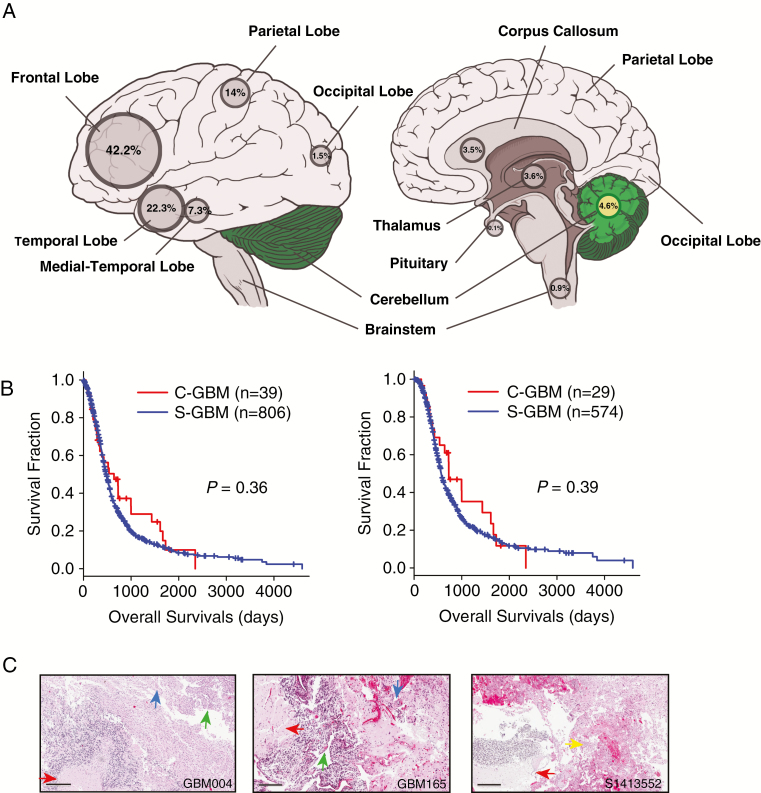
Fig. 1.
C-GBMs among 853 glioblastoma patients. (A) Location information of 853 glioblastoma patients. Among them, 39 GBMs (4.6%) were located in the cerebellum. (B) Kaplan–Meier survival plots between C-GBMs and S-GBMs for all patients (left) and temozolomide-treated patients (right). P-values were estimated by log-rank tests. (C) Histological features of C-GBM. Glioblastoma features were evident, such as tumor necrosis (red arrow), endovascular proliferation (blue arrow), hypercellularity (green arrow), and glomeruloid endovascular proliferation (yellow arrow). Scale bars, 300 μm (GBM004), 400 μm (GBM165), and 200 μm (S1413552).

