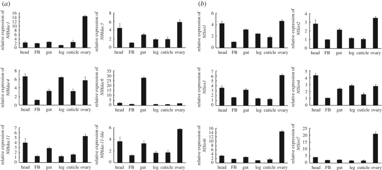
Figure 3.
Tissue distribution of the HDACs of BPH. (a) Tissue distribution of the classical HDAC family (NlHDACs). (b) Tissue distribution of the sirtuin family (NlSIRTs). Total RNAs were isolated from head, gut, fat body (FB), leg, cuticle and ovary from adult females (n = 50, 24 h after eclosion). First-strand cDNA was synthesized using random primers, and qRT-PCR was conducted using specific primers corresponding to each gene. The relative expression level was normalized by the 18S rRNA gene. Bars represent s.e.m. derived from three independent biological replicates.

