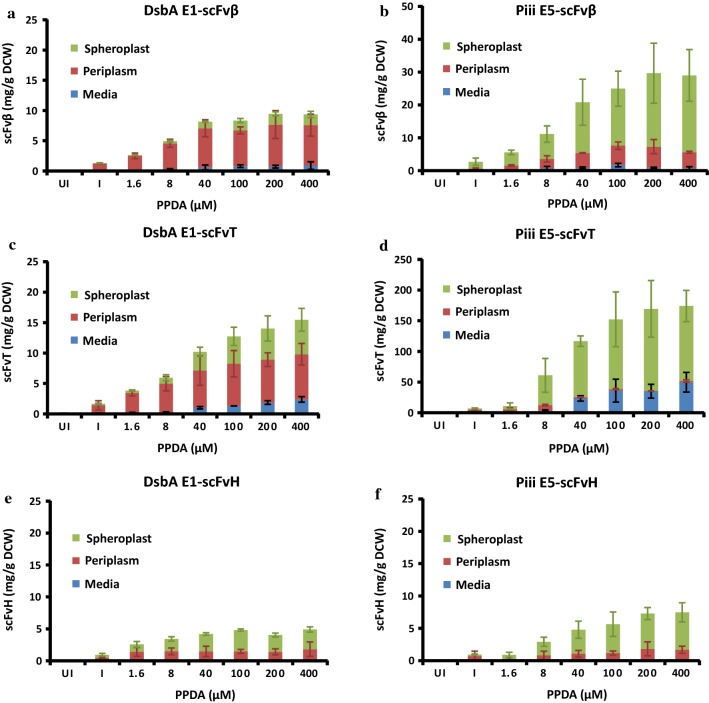
Fig. 3.
Performance of different signal peptide-scFv pDEST from shake flask expression at 30 °C, 14 h post induction. Using the DsbA E1 signal peptide sequence (a, c, e) and the Piii E5 signal peptide sequence (b, d, f) expressing scFvβ (a–b), scFvT (c–d), and scFvH (e–f). The scFv yield plotted as mg per g dry cell weight (mg/g DCW) against inducer concentrations for different scFvs, quantified from western blot analysis, from the media (M), periplasm (PP) and spheroplast (SP) fractions. Performed in the absence of inducer (UI), or with the same IPTG (I) concentration (150 μM) and increasing PPDA concentrations (1.6–400 μM). All data was taken from shaker flask expression under different inducer concentrations at 30 °C, 14 h post induction performed in biological triplicates

