Figure 3.
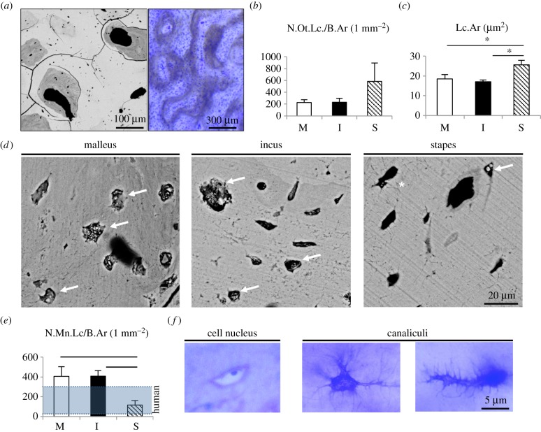
Osteocyte characteristics. (a) Left: Backscattered electron imaging of whale ossicular bone (not normalized to the grey values of qBEI) highlight osteonal structures. Lower brightness represents lower mineralization, differences in grey levels pointed to vital bone remodelling. Right: corresponding toluidine blue staining of a ground section. (b) The number of osteocyte lacunae per bone area was not different between malleus, incus and stapes. (c) In the stapes, osteocyte lacunae were enlarged compared to the malleus and incus. (d) Osteocyte lacunae were filled with calcified nanospherites (arrows). Asterisk: osteocyte canaliculi. (e) The number of mineralized lacunae was higher than in human auditory ossicles [13] in the malleus and incus and lower in the stapes. (f) Histology indicating viable osteocytes with cell nucleus (left) and branched-like osteocyte canaliculi (middle and right). *p < 0.05.

