Figure 3.

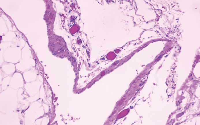
Histopathology shows that the cyst wall was composed of fibrous adipose tissue and a few lymphatic endothelial proliferations. Case 4, hematoxylin and eosin, 100 ×.

Histopathology shows that the cyst wall was composed of fibrous adipose tissue and a few lymphatic endothelial proliferations. Case 4, hematoxylin and eosin, 100 ×.