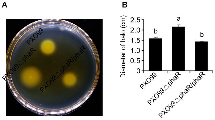
FIGURE 5.
Motility phenotypes of different strains. (A) Typical bacterial halos, formed on semisolid 0.3% agar plates after incubation at 28°C for 3 days. (B) Diameters of motility halos, formed on semisolid 0.3% agar plates. Error bars represent ±SD. Columns with different letters above were significantly different by ANOVA (p < 0.05).

