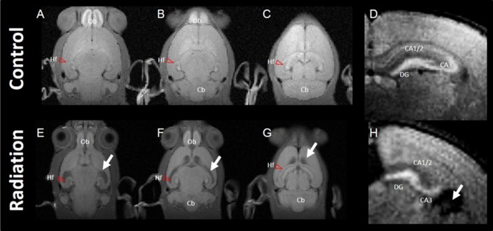
Figure 1.
Typical images of manganese-enhanced MRI (MEMRI). Top panel: MEMRI of normal rat whole brain (A–C; horizontal slice) and hippocampus (D; axial slice). Bottom panel: MEMRI of a radiation-exposed rat (E–G; horizontal slice) and hippocampus (H; axial slice). White arrows indicate ventricle dilatation in (E–H). T1 enhancement is seen around the whole hippocampal area of normal rat brain; however, T1 enhancement was different between normal and radiation-exposed rat brains, especially in dentate gyrus (DG) and CA3. Cb, cerebellum; Hf, hippocampal formation (red arrow head); Ob, olfactory bulb.

