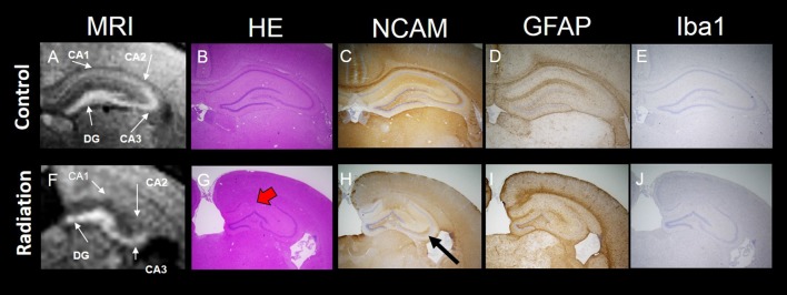
Figure 3.
MEMRI and histology of the hippocampus. Top panel: MEMRI and histology of normal rat brain (A–E). Bottom panel: MEMRI and histology of radiation-exposed rat (F–J). The cell layers in the hippocampus were subdivided into four groups: CA1, CA2, CA3 and DG. (A) MEMRI of a normal rat hippocampal region. (A,F) MEMRI of a hippocampal region. (B,G) Haematoxylin and Eosin (HE) staining. (C,H) Neural cell adhesion molecule (NCAM) staining. (D,I) Glial fibrillary acidic protein (GFAP) staining. (E,J) Ionized calcium binding adaptor molecule 1 (Iba1) staining. HE staining was more intense in normal rats than in radiation-exposed rats (B,G). Ectopic neurons were observed in radiation-exposed rats around CA1-2 (G, red arrow). NCAM immunoreactivity along the entire mossy fiber trajectory of CA3 in control rats was increased as compared to radiation-exposed rats (C,H, black arrow). The differences in positive staining were confirmed at GFAP and Iba1 only in DG between control and radiation-exposed rats (D,I). Magnification: ×40; black bar at right side in (E,J) represents 300 μm.

