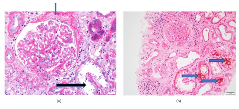
Figure 3.
Kidney biopsy histopathology II. (a) Periodic Acid Schiff (Magnification: X200) staining of glomeruli shows minimal mesangial expansion with no evidence of mesangial and endocapillary proliferation. Chronic ischemic damage is also evident by thickening of Bowman capsule membrane (blue arrow). The damaged tubules (black arrow) are also shown. (Light Microscopy.) (b) Hematoxylin and Eosin staining (Magnification: X100) shows damaged tubules with RBC casts (arrows) being seen. There is an absence of active glomerulonephritis.

