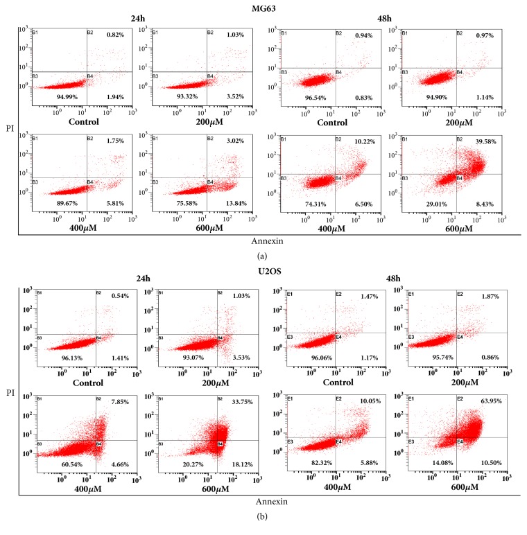
Figure 4.
Evaluation of apoptosis in MG-63 and U2OS cells by Annexin V/PI double staining assay (flow cytometry) after 24 and 48 h of treatment with different concentration of I3C. I3C induced apoptosis in a dose-and time-dependent manner in MG-63 and U2OS cells. The percentage of apoptotic cells (B2 and B4) is shown. (a) MG-63 flow cytometry dot plots. (b) U2OS flow cytometry dot plots.

