Figure 1.
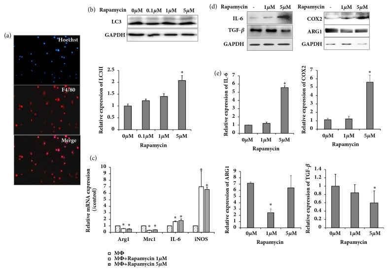
Rapamycin promoted autophagy in peritoneal macrophages and activated the M1 phenotype. (a) Peritoneal macrophages subjected to immunofluorescence staining by macrophage marker F4/80 antibody; (b) western blot showing LC3 expression in various rapamycin- (Rap-) treated groups, relative protein and GAPDH levels shown as histograms; (c) real-time PCR showing expression of Arg1, Mrc1, IL-6, and iNOS in various rapamycin-treated groups; (d) western blot showing expression of IL-6, TGF-β, COX2, and ARG1 in various rapamycin- (Rap-) treated groups; (e) relative protein and GAPDH levels shown as histograms; ∗P < .05 and ∗∗P < .01 vs. untreated controls. Results are representative of 3 independent experiments; each experiment was repeated 3 times.

