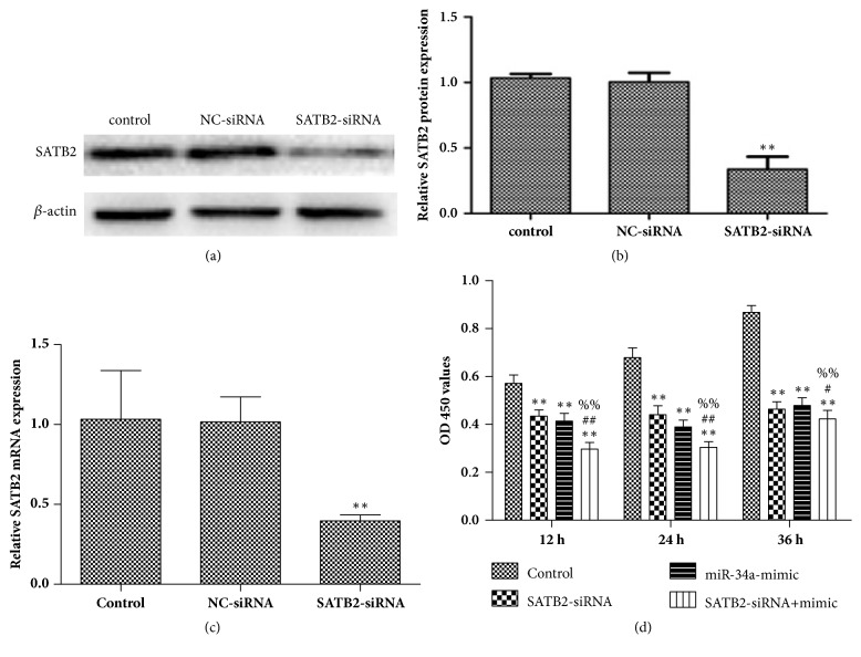
Figure 4.
miR-34a regulated cell proliferation through inhibiting SATB2. (a) Western blotting was used to detect the protein expression of SATB2 in HepG2 cells following transfection with SATB2-siRNA sequence (40 pmol/ ml) for 36 h. (b) The relative intensity of SATB2 protein was shown as a bar graph. ∗p < 0.05, versus control group. (c) RT-qPCR was applied to check the expression of SATB2 mRNA following transfection with SATB2-siRNA sequence (40 pmol/ ml) for 24 h. ∗∗p < 0.01 versus control group. (d) HepG2 cell proliferation rate was determined by a CCK-8 assay following transfection with miR-34a mimic (50 pmol/ ml) and SATB2-siRNA sequence (40 pmol/ ml) for 12, 24, and 36 h. ∗∗p < 0.01 versus control group; #p < 0.05 and ##p < 0.01 versus SATB2-siRNA group; %%p < 0.01 versus miR-34a mimic group.

