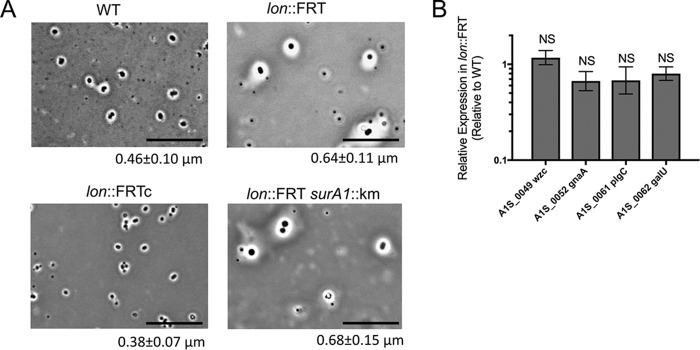
FIG 5.
Lon protease influences the cellular envelope. (A) Negative capsule staining (41) was performed on WT, lon::FRT, lon::FRTc, and lon::FRT surA1::Km strains. The white space surrounding cells is indicative of capsule. Representative microscopic images of the strains are shown. The thickness of the capsule was measured with ImageJ (NIH) (n = 100), and the mean thickness and standard deviation are provided below each image. Scale bars represent 10 μm. Staining was performed in three independent experiments. (B) Real-time qPCR was performed in biological triplicate to determine expression of the K locus in the lon::FRT and WT strains. Expression was standardized to that of 16S rRNA. Relative quantification of the K locus gene transcripts is shown, with the value for the WT strain being set to 1. Error bars represent standard deviations. An unpaired two-tailed t test was used for statistical analysis, compared to expression of the parental strain. NS, not significant (P > 0.05).

